
Uterine Cancer or Endometrial cancer : causes, symptoms, and treatment
Definition
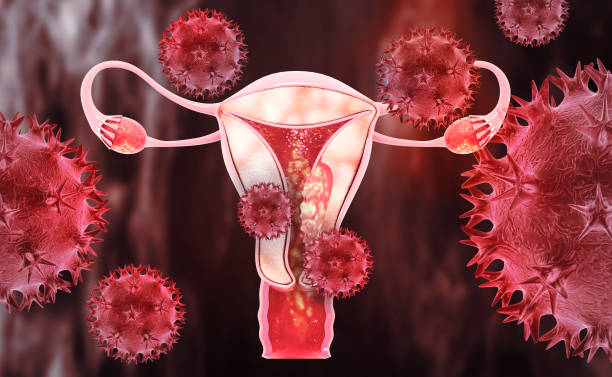
The
most prevalent cancer of the female reproductive system, uterine cancer,
develops when abnormal cells begin to grow in the uterine tissues. Through the
lymphatic and blood systems, it spreads after beginning in the uterus. Due to
the fact that the structure is a part of the uterus, cancers that develop in
each portion of the uterus have their own names, such as endometrial cancer or
cervical cancer.
The
endometrium is where the most prevalent type of uterine cancer first appears
(lining of the uterus). Uterine sarcoma is the second form of cancer that
affects the uterus. This form of uterine cancer develops in the muscle.
Types of Uterine
Cancer
There
are many different types of Uterine Cancer. Each category has different
behaviors and management requirements. We frequently request the assessment of
the results from our pathology experts because of this.
Endometrioid
adenocarcinoma: This type of uterine cancer develops in the uterine lining's
glandular cells. It may be responsible for up to 75% of all uterine
malignancies.
Endometrioid
adenocarcinoma: Frequently exhibits early detection and has a high percentage
of recovery.
Serous
adenocarcinoma :These tumors have a
higher propensity to invade nearby lymph nodes and organs. This kind of uterine
cancer is present in about 10% of cases.
Adenosquamous
cancer Both adenocarcinoma and carcinoma of the squamous cells that line the
outside of the uterus are present in this uncommon kind of uterine cancer.
Previously,
it was believed that the uncommon uterine cancer known as carcinomasarcoma was
a form of uterine sarcoma. It is currently believed to be uterine (endometrial)
cancer, though. It combines aspects of sarcoma and adenocarcinoma. The
likelihood of these tumors spreading to the lymph nodes and other organs is
very high.
Uterine
cancer risks
A
risk factor is anything that raises your likelihood of developing uterine
cancer. These consist of:
•
Obesity: Being obese doubles or quadruples your risk. Your amount of estrogen
rises as your body fat percentage rises.
•
Consuming a high-fat diet
•
Age: Women 40 and older account for more than 95% of cases of uterine cancer.
•
Tamoxifen: This breast cancer medication can increase the size of the uterine
lining. It's crucial to inform your doctor if you take tamoxifen and see
changes in your menstrual cycle or bleeding after menopause.
•
If you have a uterus, estrogen replacement treatment (ERT) without
progesterone: Pills for birth control may reduce your risk.
•
Personal /Family history of Uterine,Ovarian or Colon Cancer: This might
indicate Lynch syndrome (hereditary non-polyposis colorectal cancer or HNPCC).
Find out more about cancer syndromes that run in families.
•
Complex atypical endometrial hyperplasia, a precancerous condition that can
develop into uterine cancer if left untreated; ovarian illnesses including
polycystic ovarian syndrome (PCOS); Rarely does simple hyperplasia progress to
malignancy.
•
Diabetes.
• Never having been pregnant.
•
The number of menstrual cycles (periods) : Your risk of uterine cancer may be
increased if you began having periods before the age of 12 or experienced
menopause later in life.
•
Ovarian or breast cancer
•
Pelvic radiation for the treatment of further cancers: An individual's history
of high-dose radiation therapy in the pelvic region is the greatest risk factor
for uterine sarcoma.
Uterine
cancer does not affect everyone who has risk factors: However, it's a good idea
to address any risk factors you may have with your doctor.
Uterine
Cancer Causes
Although
the exact origin of uterine cancer is unknown, too much estrogen appears to
raise the risk.
•
The uterine lining, or endometrium, undergoes a continuous cycle of growth and
maturation during a woman's reproductive years. Progesterone promotes
maturation while estrogen promotes endometrial development. Progesterone levels
fall as a result of infertility, which results in endometrial shedding and
menstruation. High estrogen levels can cause the endometrium to expand
excessively (hyperplasia), which can turn cancerous. Endometrial cancer can
occur in people who have cancer-related syndromes, which are also present in
certain cases of uterine cancer.
Stages Of
Uterine Cancer
The
stage offers a standardized approach to describe cancer, allowing specialists
to collaborate and plan the most effective treatments. The FIGO system is used
by doctors to determine the endometrial cancer stage.
Stage
I: The cancer has not spread outside of the uterus or womb and is only present
there.
•
Stage IA: Only the endometrium or less than half of the myometrium is affected
by the malignancy.
•
Stage IB: The myometrium has been at least partially invaded by the tumor.
Stage
II: The tumor has only reached the cervical stroma and has not yet reached
other body sites, having originated in the uterus.
Stage
III: The cancer has spread outside the uterus but has not yet migrated beyond
the pelvic region.
•
Stage IIIA: Cancer has spread to the uterine serosa, fallopian tube tissue,
and/or ovaries, but not to other bodily organs.
The
tumor has reached the vagina or the area adjacent to the uterus in stage IIIB.
•
Stage IIIC1: The local pelvic lymph nodes have been affected by cancer.
•
Stage IIIC2: Cancer may have progressed to the local pelvic lymph nodes or only
to the para-aortic lymph nodes.
Stage
IV: The rectum, bladder, and/or other distant organs have been affected by
cancer metastasis.
•
Stage IVA: The bladder or rectum mucosa have been affected by cancer.
•
Stage IVB: The cancer has spread to the groin-area lymph nodes and/or distant
organs such the bones or lungs.
Signs and
Symptoms
It
is crucial that you visit a nurse, doctor, or gynecologist (a doctor who
specializes in women's health) if you have concerns about your symptoms. It is
crucial to have any symptoms investigated even though it is more likely that
they are unrelated to cancer.
•Bleeding
beyond menopause (after your periods have been absent for a year).
•Very
heavy periods, and bleeding in between periods are all signs of uterine cancer.
•
Pain in your stomach or abdomen .
•
Difficulty going to the bathroom to pass pee (wee) or discomfort when you do.
• An odd fluid or discharge from your vagina
that is watery, bloody, or foul.
If
you have any of these symptoms and they persist or seem out of the ordinary for
you, consult a doctor.
Complications
Anemia,
or a reduced red blood cell count, is the only potential side effect of
endometrial cancer symptoms. Anemia can cause symptoms such as weakness, cold
hands and/or feet, headaches, breathlessness, pale or yellow-tinged skin, chest
pain, and dizziness or lightheadedness.
This
particular type of anemia is brought on by a lack of iron in your body as a
result of blood loss. Thankfully, it can be quickly reversed by eating a diet
high in vitamins, taking iron supplements, and getting your endometrial cancer
treated, which will completely halt the bleeding. Before starting any
supplements, discuss them with your oncologist.
Diagnosis
and test
Your
medical history and current symptoms will be questioned by your doctor. You'll
also get a physical examination.
You
might undergo further tests if your doctor suspects cancer, such as:
•
A Pap test, commonly known as a pap smear, collects cells from the cervix for
laboratory analysis.
•
Ultrasound creates images of the inside of the body using sound waves. For a
better view of the uterus during a transvaginal ultrasound, the doctor inserts
a probe into the vagina. Through the cervix, sterile saline is injected during
sonohysterography into the uterus. This adds further information.
•
Tissue samples from the uterus are taken during a biopsy for laboratory
investigation. A biopsy is most often the only
certain way to tell if cancer is present.
•
A procedure called dilation and curettage (D&C) is used to take tissue
samples from the uterus. It is frequently combined with a hysteroscopy so the
surgeon may see the uterine lining while doing the surgery.
If
cancer is found, one or more of the tests listed below can determine whether it
has spread:
•
Your uterus, lymph nodes, and other abdominal tissues can be seen in great
clarity in body MRI images. In order to make lymph nodes and other tissues more
visible on an x-ray, your doctor may inject you with contrast material. MRI is
helpful for planning treatments and staging diseases.
•
Your chest, belly, or pelvis can all be seen in great detail thanks to a body
CT scan. In order to make lymph nodes and other tissues more visible on an
x-ray, your doctor may inject you with contrast material. CT scans can detect
cancer in the lungs, lymph nodes, uterus, and other places.
•
A chest x-ray creates images of the lungs on film.
•
A PET scan makes use of a little amount of radioactive substance to assist in
determining the severity of your cancer. To create unique viewpoints, PET scans
can be placed on CT or MRI images. These perspectives may result in more
accurate diagnosis.
Treatment
One
or more therapies, such as laparoscopic surgery, radiation therapy,
chemotherapy, and hormone therapy, are used to treat uterine cancer. Depending
on the tumor risk factors, 10-15% of patients may require adjuvant radiotherapy
or radiotherapy combined with chemotherapy.
Surgery:
The act of removing a tumor and its surrounding tissue through an operation.
Typical
surgical techniques include:
•
Hysterectomy: Using a laparoscope, the uterus and cervix are removed during
this treatment. For patients who have undergone menopause, the surgeon may also
conduct a bilateral salpingo-oophorectomy, in which both fallopian tubes and
ovaries are removed. The procedure can be basic or drastic.
•
Lymphadenectomy: Lymph nodes close to the tumor are removed during this
surgery.
Radiation
therapy, which uses x-rays to damage or kill cancer cells, is frequently
employed as a supplemental therapy to lower the risk of cancer recurrence. If
you are not well enough for surgery, it might be suggested as your primary
course of treatment. Radiation therapy can be administered externally, using a
machine to target the cancer and surrounding tissue, or internally
(brachytherapy), using radioactive material inserted into tiny tubes and placed
close to the tumour.
It
is crucial to discuss your concerns with your treatment team before the start
of treatment if you are worried about how the treatment will effect your
fertility. Radiation therapy to the pelvic region has the potential to produce
menopause.
Chemotherapy:
Chemotherapy is used to treat some types of uterine cancer, as well as when the
disease recurs after radiotherapy or surgery or if it does not respond to
hormone therapy. Both symptom relief and cancer control are possible with it.
Typically, a medicine is administered by injecting it into a vein
(intravenously). The course and duration of the chemotherapy treatment will be
described by the doctor.
Hormone
treatment It is typically used if cancer has spread or if it has returned
(recurred). In rare cases, it is utilized in place of surgery. The primary
hormone therapy for uterine cancer patients is progesterone, which can be
administered orally or intravenously by a doctor or nurse. It aids in symptom
control and the reduction of some malignancies.
Surgery
alone can cure more than 80% of endometrial cancer cases. Depending on your
condition, the doctor may urge you to stay in the hospital for 3-5 days.
Prevention
of Uterine Cancer
Uterine
carcinoma cannot be entirely avoided. Generally speaking, prevention methods
involve reducing risk variables that are within your power.
·
Consuming a healthy diet
·
Maintaining a healthy weight
·
Managing other medical conditions like diabetes,
·
Quitting smoking
·
Discussing your risk with your doctor before starting hormone
replacement therapy (HRT) for menopause or other conditions.
·
Attending routine physical exams and OBGYN appointments are some
examples of what this entails.


